http://radiopaedia.org/articles/lissencephaly-pachygyria-spectrum-1/revisions/21
Lissencephaly – pachygyria spectrum
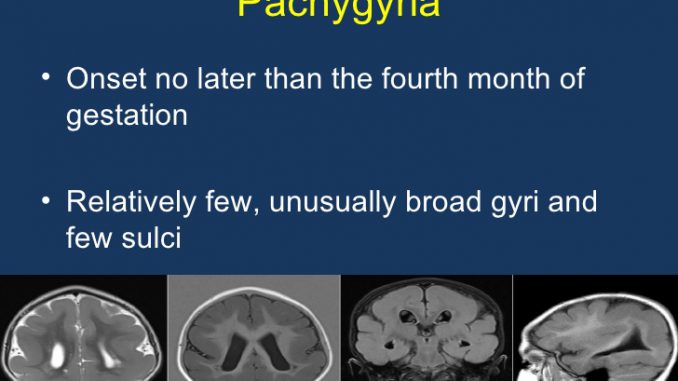
Lissencephaly (also known as agyria) is a congenital cerebral malformation and it lies along a continuum of pachygyria; as such the term lissencephaly – pachygyira spectum is often used. It a on of many disorders of cortical formation 5, which results in a smooth cortical surface lacking the normal gyral convolutions.
Lissencephaly – pachygyria can be further divided into types I (classic) and type II, which differ in clinical presentation, underlying genetic abnormalities, and both microscopic and macroscopic (including imaging) appearances 2,6.
As such they are discussed separately:
type I (classic) lissencephaly
type II (cobble stone complex) lissencephaly
Clinical presentation
Type I (classic) lissencephaly typically presents with marked hypotonia and paucity of movement.
Type II lissencephaly is associated with muscular dystrophy-like syndromes.
— Walker-Warburg syndrome
— Fukuyama syndrome: predominantly reported in Japanese populations
— muscle-eye-brain (MEB) disease: predominantly reported in Finnish populations
Pathology
Sub types
classic lissencephalies (type I): small Sylvian fissure with an “hour glass” or “figure-8” cerebral configuration arising as a result of arrest or delay of neuronal delay between 9 – 16 weeks gestation 2,3. These are part of the spectrum which encompases agyria / pachygyria, although increasingly individual sub-conditions have been isolated to individual gene mutations:
lissencephaly due to LIS1 gene mutation, which subdivides into:
type I isolated lissencephaly
Miller-Dieker syndrome (MDS)
lissencephaly due to doublecortin (DCX) gene mutation
lissencephaly, type I, isolated, without a known genetic defect.
cobblestone lissencephaly (type II): usually associated with muscular dystrophy syndromes and results from over-migration of neurons through the cortex. Similar changes are seen in the cerebellum. The cortex is less thick than in type I
lissencephaly X-linked with agenesis of the corpus callosum
lissencephaly with cerebellar hypoplasia, including
Norman-Roberts syndrome (mutation of reelin gene)
microlissencephaly (lissencephaly and microcephaly)
Radiographic features
Although lissencephaly can be identified on all cross-sectional modalities (antenatal and neonatal ultrasound, CT and MRI), MRI is the modality of choice to fully characterise the abnormalities.
MRI
Type I and type II lissencephaly demonstrate different macroscopic and imaging appearances.
Type I (classic) lissencephaly can apear as the classic hour glass or figure-8 appearance or with a few poorly formed gyri (pachygyria).
Type II lissencephaly on the other hand has a microlobulated surface referred to cobblestone complex.
Differential diagnosis
early fetal brain : prior to 22 weeks : lacks gyral patterns


