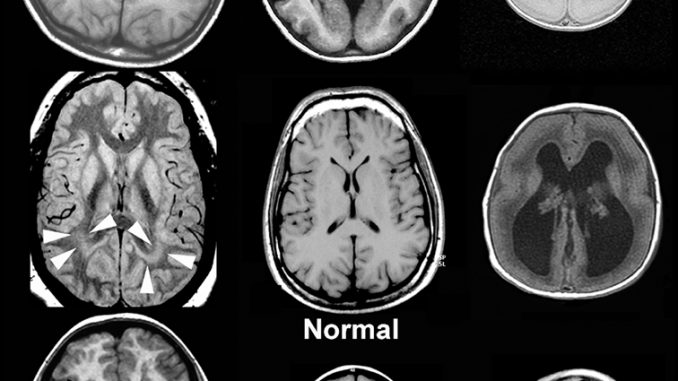
Malformations of Cortical Development: Diagnostic Accuracy of Fetal MR Imaging
Orit A. Glenn, MD, Addison A. Cuneo, BS, A. James Barkovich, MD, Zary Hashemi, MD, Agnes I. Bartha, MD and Duan Xu, PhD + Author Affiliations
From the Department of Radiology and Biomedical Imaging, Neuroradiology Section, University of California, San Francisco, 505 Parnassus Ave, Box 0628, San Francisco, CA 94143-0628.
Abstract
Purpose:
To determine the diagnostic accuracy of fetal magnetic resonance (MR) imaging for malformations of cortical development by using postnatal MR imaging as reference standard.
Materials and Methods:
Eighty-one patients who had undergone fetal and postnatal MR imaging of the brain were identified in this institutional review board–approved, HIPAA-compliant study. Images were retrospectively reviewed in consensus by two pediatric neuroradiologists who were blinded to clinical information. Sensitivity and specificity were calculated according to retrospective review of the images and clinical reports for fetal MR images. The Fisher exact test was used to compare results for fetuses imaged before and after 24 gestational weeks and for image review versus clinical reports for fetal MR images.
Results:
Median gestational age at fetal MR imaging was 25.0 weeks (range, 19.71–38.14 weeks). Postnatal MR imaging depicted 13 cases of polymicrogyria, three cases of schizencephaly, and 15 cases of periventricular nodular heterotopia. Sensitivity and specificity of fetal MR imaging were 85% and 100%, respectively, for polymicrogyria; 100% each for schizencephaly; and 73% and 92%, respectively, for heterotopia. When heterotopia was seen in two planes, specificity was 100% and sensitivity was 67%. Sensitivity for heterotopia decreased to 44% for fetuses younger than 24 weeks. According to reports for fetal MR images, prospective sensitivity and specificity, respectively, were 85% and 99% for polymicrogyria, 100% and 99% for schizencephaly, and 40% and 91% for heterotopia.
Conclusion:
Fetal MR imaging had the highest sensitivity for polymicrogyria and schizencephaly. Specificity was 100% for all cortical malformations when the abnormality was seen in two planes. Sensitivity for heterotopia was lower for fetuses younger than 24 weeks. Knowledge of the gestational age is important, especially for counseling patients about heterotopia.
© RSNA, 2012
Footnotes
Received December 21, 2010; revision requested February 11, 2011; final revision received August 8; accepted August 26; final version accepted December 13.
Funding: This research was supported by the National Institutes of Health (grant K23 NS52506).
The contents of this study are solely the responsibility of the authors and do not necessarily represent the official views of the National Institutes of Health.
Abbreviations:
CI =
confidence interval
PVNH =
periventricular nodular heterotopia
SE =
spin echo
Author contributions:
Guarantor of integrity of entire study, O.A.G.; study concepts/study design or data acquisition or data analysis/interpretation, all authors; manuscript drafting or manuscript revision for important intellectual content, all authors; approval of final version of submitted manuscript, all authors; literature research, O.A.G., A.J.B., A.I.B., D.X.; clinical studies, O.A.G., A.A.C., A.J.B., Z.H., D.X.; statistical analysis, O.A.G., A.A.C., Z.H., A.I.B., D.X.; and manuscript editing, O.A.G., A.J.B., D.X.
Address correspondence to
O.A.G. (e-mail: Orit.Glenn@ucsf.edu).


